An in-depth explanation of how laboratory exercises demonstrate the principle of phage typing, including mechanisms, methodology, and real-world applications.
Introduction
Understanding how microorganisms are identified and differentiated is a cornerstone of microbiology. One of the most fascinating and precise techniques used for this purpose is phage typing. This method relies on the specificity of bacteriophagesviruses that infect bacteriato distinguish between bacterial strains. In many laboratory settings, students perform exercises that simulate or directly demonstrate this concept, allowing them to observe bacterial susceptibility patterns and understand how phage typing works in practice.
This article explores in detail how a typical laboratory exercise demonstrates the principle of phage typing, breaking down the science behind it, the steps involved, and its significance in research and clinical diagnostics.
What is Phage Typing?
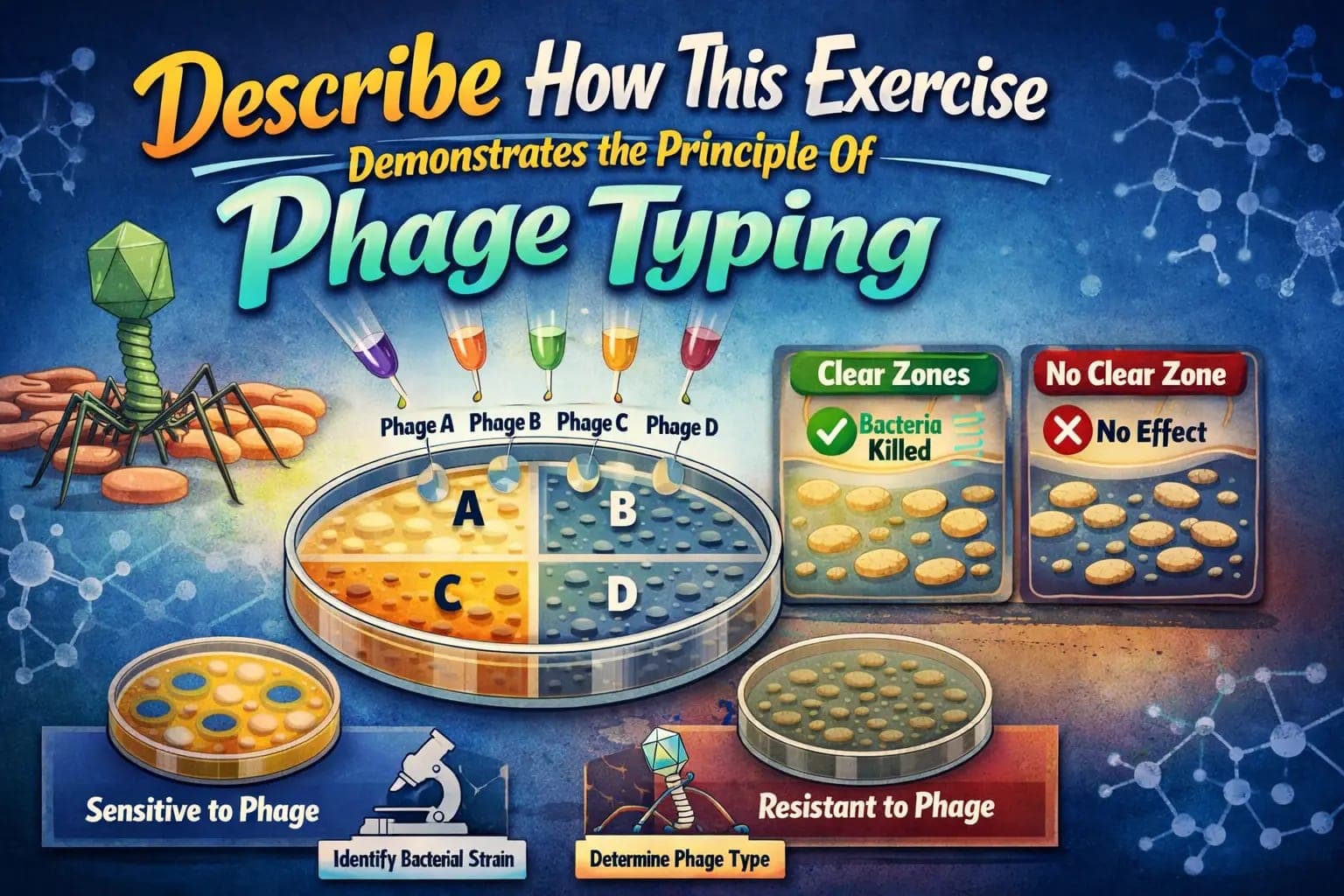
Phage typing is a microbiological technique used to identify and differentiate bacterial strains based on their susceptibility to specific bacteriophages. Each bacteriophage is highly selective and can infect only certain bacteria. By exposing bacteria to a panel of phages, scientists can observe patterns of lysis (destruction of bacterial cells) and use these patterns to classify the bacteria.
This method has historically been used to track outbreaks of infectious diseases, such as typhoid fever and staphylococcal infections.
The Principle Behind Phage Typing
The principle of phage typing is based on host specificity. Bacteriophages recognize and bind to specific receptors on the surface of bacterial cells. If a bacterium possesses the correct receptor, the phage attaches, injects its genetic material, and ultimately causes the bacterial cell to lyse.
If the receptor is absent or altered, the phage cannot infect the bacterium. This specificity allows microbiologists to create a "phage sensitivity profile" for each bacterial strain.
Overview of the Laboratory Exercise
A typical exercise designed to demonstrate phage typing involves the following components:
- A bacterial culture (often a known strain)
- A set of bacteriophages
- Nutrient agar plates
- Sterile tools for inoculation
The goal of the exercise is to expose the bacterial culture to different phages and observe which ones cause lysis.
Step-by-Step Procedure of the Exercise
1. Preparation of Bacterial Lawn
Students begin by spreading a uniform layer of bacterial culture across an agar plate. This creates what is known as a bacterial lawn, which provides a continuous surface for observing phage activity.
2. Application of Phages
Small drops of different bacteriophage solutions are placed onto marked sections of the agar surface. Each drop contains a specific type of phage.
3. Incubation
The plate is incubated at an optimal temperature (usually 35–37°C) for 18–24 hours. During this time, phages interact with the bacterial cells.
4. Observation of Results
After incubation, students examine the plate for zones of clearance, also known as plaques. These clear areas indicate where bacterial cells have been lysed by phages.
How the Exercise Demonstrates the Principle of Phage Typing
1. Visualization of Host Specificity
One of the most direct ways this exercise demonstrates the principle of phage typing is through visible evidence of host specificity. Not all phage spots produce plaques. Only those phages capable of infecting the bacteria will create clear zones.
This clearly shows that:
- Phages are selective
- Bacteria differ in susceptibility
- Interactions are highly specific
2. Creation of a Lysis Pattern
Each bacterial strain produces a unique pattern of lysis depending on which phages affect it. In the exercise, students can record which phage spots show clearing and which do not.
This pattern mimics real-world phage typing, where such profiles are used to identify bacterial strains.
3. Understanding Bacterial Diversity
Even closely related bacterial strains can show different susceptibility patterns. This exercise helps students understand that bacterial populations are not uniform and that subtle genetic differences can influence phage infection.
4. Demonstration of Virus-Host Interaction
The experiment highlights the biological interaction between viruses and bacteria. Students observe firsthand how a virus can infect and destroy a host cell, reinforcing theoretical knowledge with practical evidence.
5. Linking Theory to Practical Application
Phage typing is often discussed in textbooks, but this exercise bridges the gap between theory and practice. By performing the steps themselves, students gain a deeper understanding of how microbiologists use this technique in laboratories.
Scientific Concepts Reinforced by the Exercise
A. Specificity of Biological Interactions
The exercise demonstrates that biological interactions are highly specific. Just as enzymes bind to specific substrates, bacteriophages bind to specific bacterial receptors.
B. Genetic Variation in Bacteria
Differences in phage susceptibility often arise from genetic variation. This reinforces the concept that even small genetic changes can have significant functional consequences.
C. Viral Replication and Lysis
Students observe the end result of viral replicationthe destruction of bacterial cells. This provides insight into the lytic cycle of bacteriophages.
D. Microbial Identification Techniques
The exercise introduces students to one of many methods used to identify and classify microorganisms.
Real-World Applications of Phage Typing
1. Epidemiology
Phage typing has been used to trace the source of bacterial outbreaks. By comparing phage sensitivity patterns, scientists can determine whether infections originate from the same strain.
2. Clinical Diagnostics
In clinical settings, phage typing can help identify pathogenic bacteria and guide treatment decisions.
3. Research and Biotechnology
Researchers use phage typing to study bacterial genetics, evolution, and resistance mechanisms.
4. Food Safety
The technique is also applied in monitoring contamination in food production systems.
Advantages of Phage Typing
- High specificity
- Relatively simple methodology
- Useful for strain differentiation
- Cost-effective in basic laboratory setups
Limitations of Phage Typing
While the exercise demonstrates the principle effectively, it also highlights certain limitations:
- Not all bacteria are typeable using phages
- Requires a well-maintained phage library
- Results can sometimes be ambiguous
- Being gradually replaced by molecular techniques like PCR and genome sequencing
Modern Alternatives and Complementary Techniques
Although phage typing is still valuable, modern microbiology often uses advanced methods such as:
- DNA sequencing
- Polymerase Chain Reaction (PCR)
- Pulsed-field gel electrophoresis (PFGE)
These methods provide higher accuracy and reproducibility, but they also require more sophisticated equipment.
Educational Importance of the Exercise
From a teaching perspective, this exercise is অত্যন্ত valuable because it:
- Encourages hands-on learning
- Reinforces theoretical concepts
- Develops laboratory skills
- Enhances critical thinking and observation
Students not only learn about phage typing but also gain experience in sterile techniques, data recording, and result interpretation.
Conclusion
The laboratory exercise designed to demonstrate the principle of phage typing is a powerful educational tool. By observing the interaction between bacteriophages and bacteria, students gain a clear understanding of host specificity, viral infection, and microbial diversity.
Through the formation of plaques and the creation of lysis patterns, the exercise brings the concept of phage typing to life. It effectively shows how bacteria can be differentiated based on their susceptibility to specific phages, mirroring real-world applications in epidemiology, diagnostics, and research.
While newer molecular methods are becoming more prevalent, the fundamental principles illustrated by this exercise remain essential to the study of microbiology. Ultimately, it serves as a perfect example of how simple experiments can reveal complex biological truths and inspire deeper scientific inquiry.